Cruciate Ligament Surgery
What is a Cruciate Ligament Injury?
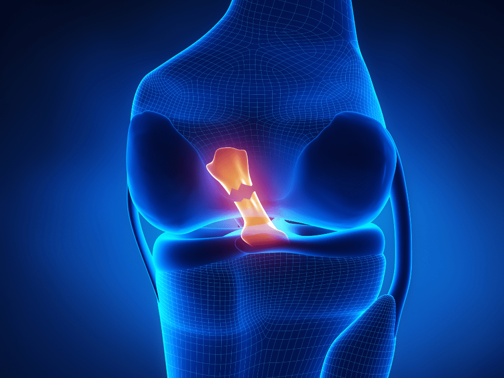
Cruciate ligament injuries are one of the most common orthopaedic problems in pets, particularly in dogs. These injuries involve the tearing or rupture of the cranial cruciate ligament (CCL) in the knee joint. This ligament plays a critical role in stabilising the knee, and when it is damaged, pets can experience significant pain, lameness, and a decreased quality of life.
What are the causes?
Cruciate ligament injuries can result from a variety of factors, including:
- Trauma: Sudden twisting or turning of the knee, often during play or exercise.
- Obesity: Excess weight puts additional stress on the knee joint, increasing the likelihood of ligament damage.
- Genetics: Certain breeds are predisposed to cruciate ligament injuries, such as Labrador Retrievers, Rottweilers, and Bulldogs.
- Age: Older pets may experience degenerative changes in the ligament, making it more prone to injury.
Signs and Symptoms
If your pet is suffering from a cruciate ligament injury, you may notice the following signs:
- Sudden lameness in one or both hind legs
- Difficulty rising, walking, or running
- Swelling around the knee joint
- Reluctance to bear weight on the affected leg
- Pain or discomfort when the knee is touched
If you observe any of these symptoms in your pet, it’s important to seek veterinary care as soon as possible.

ZLIG - ACL Replacement
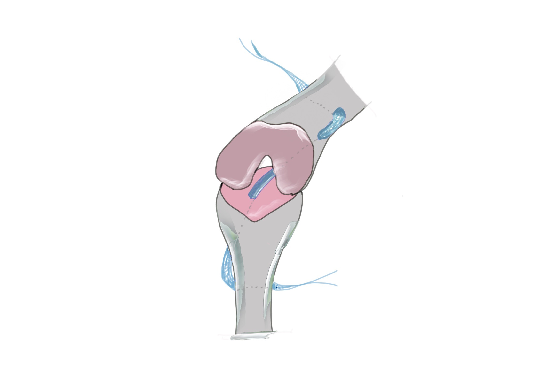
Zlig is an innovative artificial implant designed to replace a torn Anterior Cruciate Ligament (ACL) in dogs. Unlike other procedures that adjust knee function to compensate for an ACL tear, Zlig directly stabilises the ACL. This method, standard in human medicine, is increasingly recommended for dogs due to advancements that reduce complications and enhance joint strength and stability.
How does a Zlig Implant work?
Constructed from materials similar to those in the LARS procedure for humans, the Zlig implant serves as a durable ACL substitute. It integrates seamlessly into the joint, promoting fibroblast growth to mimic a natural ACL. The implant is secured with four screws in drilled bone tunnels, minimizing infection and fracture risks as no bone is removed during the process.
What are the benefits?
-
Direct ACL Stabilization: Unlike other procedures that alter knee function, Zlig directly stabilises the ACL, leading to a more natural recovery.
-
Reduced Complications: Advancements in the procedure have led to fewer health complications, including minimized risks of infection and fractures.
-
Enhanced Longevity: The Zlig implant promotes better long-term joint stability, potentially reducing the risk of arthritis compared to other ACL treatments.
-
Quick Recovery: Most dogs can start using their leg soon after surgery and return to normal activities within a couple of months
Lateral Suture Technique (LS)
What is the Lateral Suture Technique?
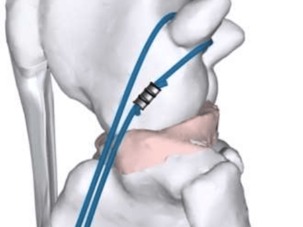
The Lateral Suture Technique, often referred to as Extracapsular Repair, is a preferred approach for smaller dogs and cats. This surgical method involves the placement of a robust suture material outside the knee joint, effectively emulating the role of the damaged cruciate ligament. By stabilizing the knee, the suture allows for the formation of scar tissue around the joint, ensuring long-lasting stability and improved functionality.
How does Lateral Suture Technique work?
The Lateral Suture Technique is a surgical procedure used to stabilize the knee in pets with a torn cranial cruciate ligament (CCL), commonly in small to medium-sized dogs. During the surgery, a strong suture is placed outside the knee joint, mimicking the function of the damaged ligament and restoring stability. Over time, scar tissue forms around the suture, providing long-term joint stability. This method is less invasive and more affordable than other options, though it may be less effective in larger, more active pets.
What are the benefits?
- Less Invasive: The procedure is less invasive compared to other surgical options like TPLO or TTA, leading to reduced surgical trauma.
- Cost-Effective: It is generally more affordable, making it a viable option for pet owners on a budget.
- Shorter Recovery Time: Pets typically experience a quicker recovery compared to more complex surgeries, allowing them to return to normal activities sooner.
- Effective for Small to Medium-Sized Pets: The technique is particularly effective for smaller dogs and cats, offering a reliable solution for pets that aren’t excessively active.
- Immediate Stability: The suture provides immediate stabilization of the knee joint, helping to reduce pain and improve mobility right after surgery.
- Simpler Procedure: The surgery is straightforward, reducing the risk of complications during the procedure.
Modified Maquet Procedure (MMP)
What is the Modified Maquet Procedure?
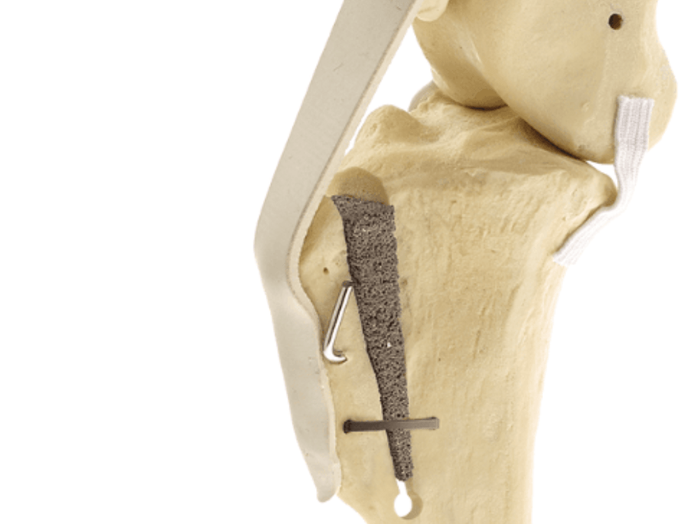
- The Modified Maquet Procedure (MMP) is a surgical technique used to treat cranial cruciate ligament (CCL) injuries in dogs. It’s an evolution of the Tibial Tuberosity Advancement (TTA) surgery, designed to stabilise the knee joint by altering its biomechanics.
How does Lateral Suture Technique work?
- Tibial Tuberosity Advancement: MMP involves advancing the tibial tuberosity, which is the part of the shin bone where the patellar ligament attaches. By altering the angle of the knee joint, this advancement neutralises the forces that cause instability, effectively making the damaged cruciate ligament unnecessary for knee stability.
- Use of a Wedge-Shaped Cage: A wedge-shaped titanium cage is inserted into a cut made in the tibial tuberosity. This cage holds the advancement in place, providing precise and stable support for the bone as it heals. The cage allows for a more accurate and consistent correction compared to traditional methods.
- Bone Healing: Over time, the bone heals around the titanium cage, which stabilises the knee without relying on the damaged ligament. This surgical change in the knee’s biomechanics reduces the risk of re-injury and provides long-term stability.
What are the benefits?
- Enhanced Stability: The titanium cage provides precise and stable knee joint support.
- Faster Recovery: MMP typically promotes quicker healing compared to other techniques.
- Minimally Invasive: The procedure is less invasive, leading to less pain and a shorter recovery period.
- Long-Term Success: The biomechanical changes reduce the risk of re-injury, offering lasting stability.
- Effective for Active Dogs: Particularly beneficial for large or highly active dogs needing robust knee support.
Cranial Closing Wedge Osteotomy (CCWO)
What is a CCWO procedure?
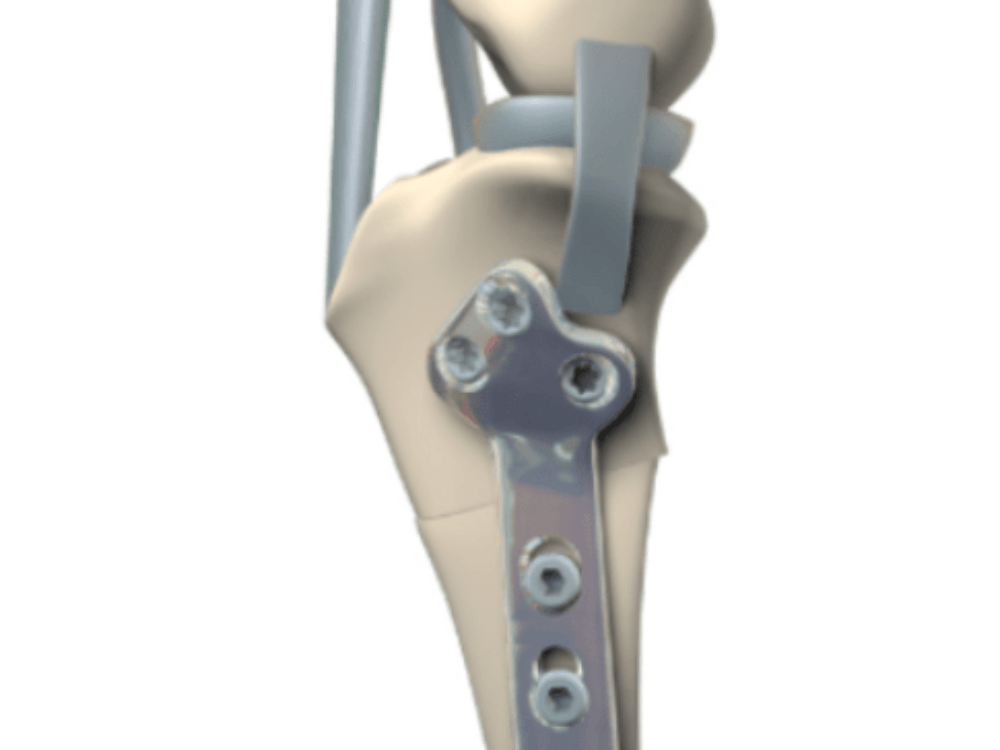
Cranial Closing Wedge Osteotomy (CCWO) is a surgical procedure used to treat cranial cruciate ligament (CCL) injuries in dogs. It is an alternative to other techniques like TPLO (Tibial Plateau Leveling Osteotomy), particularly in cases where the anatomy of the dog’s tibia makes TPLO less suitable.
How does a CCWO work?
-
Wedge-Shaped Bone Removal: During CCWO, the surgeon removes a wedge-shaped piece of bone from the top of the tibia (shin bone). This cut is made near the knee joint, at the front (cranial) aspect of the tibia.
-
Bone Realignment: After removing the wedge, the tibia is realigned to change the angle of the tibial plateau, which is the top surface of the tibia that forms part of the knee joint. The new angle helps to neutralize the abnormal forces in the knee caused by the damaged CCL, stabilising the joint without the need for the ligament.
-
Bone Stabilization: The tibia is then stabilised using plates and screws to hold the bone in its new position. As the bone heals, the knee becomes stable and the pet can gradually return to normal activity.
What are the benefits?
- Effective for Certain Anatomies: CCWO is particularly beneficial for dogs with a steep tibial plateau angle, where other surgeries like TPLO might be less effective.
- Improved Joint Stability: By altering the tibial plateau angle, CCWO provides long-term joint stability, reducing pain and improving mobility.
- Durability: The procedure is highly effective in preventing future knee injuries, especially in active dogs.
- Adaptable Technique: CCWO can be tailored to the specific anatomy of the dog, offering a customised approach to CCL repair.
Tibial Plateau Levelling Osteotomy (TPLO)
What is A TPLO procedure?
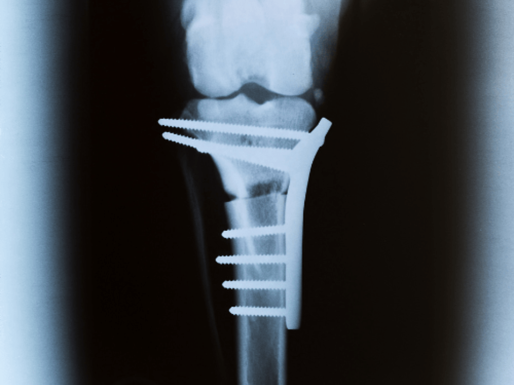
TPLO is a more advanced surgical procedure that is often recommended for larger dogs or those with high activity levels. During a TPLO, the surgeon cuts and rotates the tibial plateau (the top part of the shin bone) to change the angle of the knee joint. This adjustment stabilizes the knee without relying on the cruciate ligament.
How does a TPLO work?
- Tibial Plateau Leveling Osteotomy (TPLO) stabilizes a dog’s knee after a cranial cruciate ligament (CCL) injury by altering the angle of the tibial plateau, the top part of the shin bone. The surgeon cuts and rotates this bone to a more level position, which changes the knee’s mechanics, making the damaged ligament unnecessary for stability. The bone is then secured with a metal plate and screws, allowing it to heal in the new alignment. TPLO is especially effective for large or active dogs, offering long-term joint stability and reducing the risk of re-injury.
What are the benefits?
- Enhanced Joint Stability: TPLO effectively stabilizes the knee by altering its biomechanics, reducing pain and improving mobility.
- Reduced Risk of Re-Injury: The procedure minimises the likelihood of future knee injuries by eliminating the need for the damaged ligament.
- Suitable for Large and Active Dogs: TPLO is particularly effective for larger, more active dogs, who require a strong and stable knee.
- Quick Recovery: Many dogs can return to normal activities relatively quickly after TPLO compared to other surgical options.
- Long-Term Success: The procedure has a high success rate, providing lasting relief and allowing dogs to maintain an active lifestyle.
FAQ
Are there any risks associated with Cruciate Ligament Surgery?
As with any surgery, there are risks, including infection, bleeding, or complications related to anesthesia. Specific risks for cruciate ligament surgery may include suture failure, incomplete healing, or arthritis development. Our Vet will discuss potential risks and how they are managed.
How can I prevent Cruciate Ligament injuries?
Maintaining a healthy weight, providing regular exercise, and avoiding activities that put excessive stress on the joints can help reduce the risk of future injuries. Discuss with your veterinarian about any specific preventive measures suitable for your pet.
Book an Appointment:
If you’re concerned about your pet’s cruciate ligament health, don’t wait for the symptoms to worsen; take the proactive step of booking a consultation with our experienced team at Warren House Veterinary Centre.

