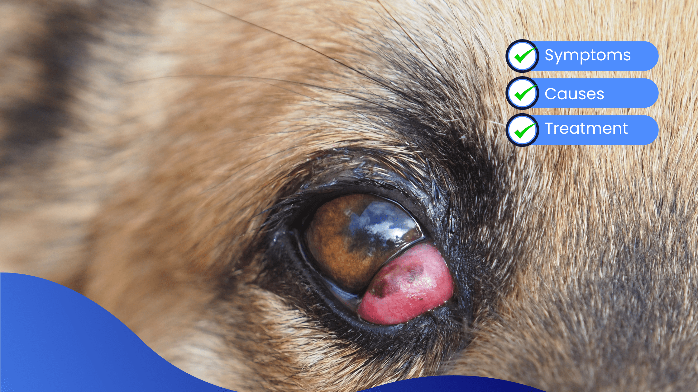
Cherry eye surgery in dogs is a procedure performed to correct the condition known as cherry eye, where the gland of the third eyelid (nictitans gland) protrudes or prolapses from its normal position.
Did you know? The third eyelid, or nictitating membrane, is a protective membrane located in the inner corner of the eye. It contains a tear gland that contributes to the production of tears.
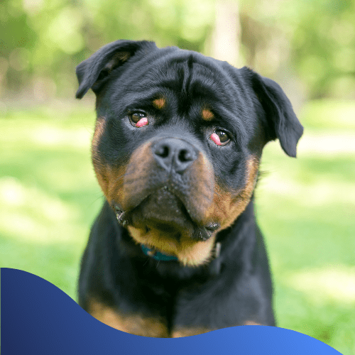
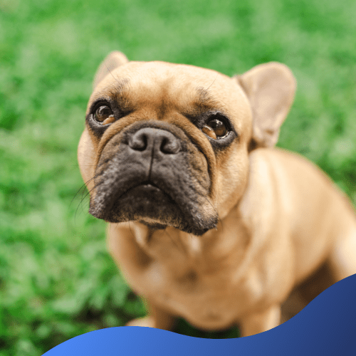
Cherry eye occurs when the gland in the third eyelid slips out of its normal position and becomes swollen, appearing as a red or pink mass resembling a cherry. This condition is more common in certain breeds, such as Bulldogs, Beagles, Cocker Spaniels, and Boston Terriers, but it can affect any breed.
Common Symptoms Of Cherry Eye:
- Visible Red or Pink Mass – The prolapsed gland is usually noticeable in the inner corner of the eye.
- Excessive Tearing – Increased tearing or eye discharge may occur.
- Blinking or Squinting – Dogs with cherry eye may blink or squint due to discomfort.
Potential Causes Of Cherry Eye
- Genetics – Evidence suggests a genetic predisposition to cherry eye, and certain breeds are more prone to this condition.
- Anatomical Factors – Dogs with a shallow eye socket or a more prominent bulging eye may be at a higher risk of developing cherry eye.
- Connective Tissue Weakness – That supports the gland in the third eyelid can lead to its prolapse.
Cherry eye often becomes more common in young dogs, typically under the age of two.
Treatment Options For Cherry Eye
- Medical Management – Vets can attempt to resolve the issue with medications to reduce inflammation, this depends on the severity of the condition.
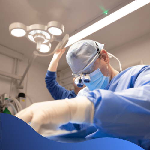
- Surgery – Correction is strongly recommended to reposition the gland and prevent recurrence.
If left untreated, cherry eye can lead to complications such as dry eye and other eye problems.
Surgery Options
Gland Replacement (Pocket Technique)
This method involves creating a small pocket or pouch within the conjunctiva and placing the prolapsed gland into this pocket. The tissue is then sutured to secure the gland in its normal position.
Advantage: It aims to keep the gland in its location while minimising the risk of injury to the gland and surrounding tissues.
Gland Resection (Excision Technique)
If the gland is severely damaged or the pocket technique fails due to the anatomy of the dog’s eye, the prolapsed gland may be partially removed (excised). This is less common.
Advantage: This method may prevent further recurrence but it may lead to reduced tear production.
Surgical Procedure:
- Preoperative Consult– The vet will perform a thorough examination to assess the extent of the Cherry Eye and rule out any underlying eye issues.
- Anesthesia – Your dog will be placed under general anaesthesia to make sure they remain, calm and comfortable during the surgery.
- Cherry Eye Repositioning – Adapting the pocket or excision technique depending on the severity of the Cherry Eye.
- Postoperative Care – After the surgery, your dog will be closely monitored as they recover from the anaesthesia. Your vet may prescribe medication, such as antibiotics or anti-inflammatory drugs, to aid in the healing process.
- Postoperative Consult – Your vet will schedule a follow-up appointment and make certain the cherry eye has been successfully corrected.
Benefits Of Cherry Eye Surgery
Relief from discomfort – Cherry eye surgery helps alleviate discomfort associated with the prolapsed gland, reducing irritation and the risk of secondary complications. The surgery will also restore the normal appearance of the eye by eliminating the red, swollen mass caused by the protruding gland.
If you notice any abnormalities in your dog’s eyes or suspect your dog has cherry eye, it is vital you contact us. We will assess the severity of the condition and recommend the appropriate treatment. Early intervention can prevent complications and contribute to your dog’s overall eye health and well-being.
We hope you have found this information helpful, until next time…