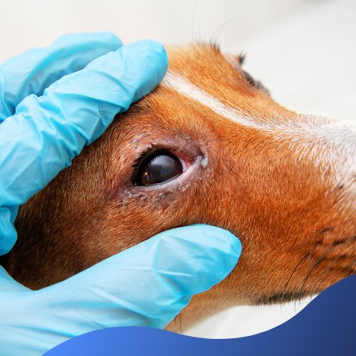
Various corneal conditions can affect dogs, ranging from mild irritations to more serious issues. Here are some common corneal conditions seen in dogs, the symptoms to look out for, and what the treatments are.
Let’s have a look at the conditions:
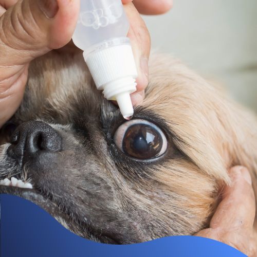
Corneal Abrasion
A scratch or abrasion to the corneal surface.
Causes: Trauma from foreign objects, scratches, or other injuries.
Symptoms:
- Squinting
- Tearing
- Redness
- Sensitivity to light
Treatment:
Antibiotics and pain management
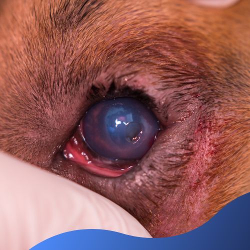
Corneal Ulcer
A sore or wound on the corneal surface.
Causes: Trauma, infections (bacterial, viral, or fungal), dry eye, or abnormalities in eyelid conformation.
Symptoms:
- Squinting
- Tearing
- Redness
- Sensitivity to light
- Cloudiness or opacity in the affected eye.
Treatment:
Antibiotics, lubricating eye drops, medications for pain and inflammation. In severe cases, surgical intervention may be required, such as Diamond Burr.
Keratoconjunctivitis Sicca (Dry Eye)
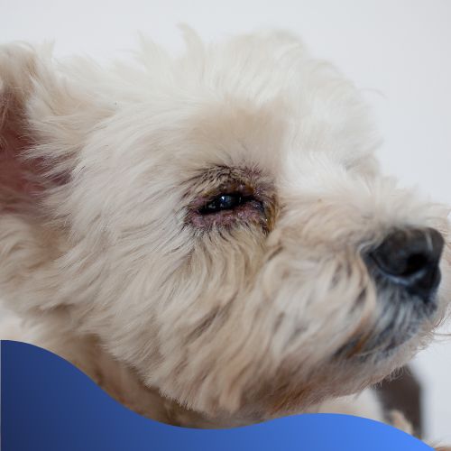
Insufficient tear production leads to dryness of the cornea.
Causes: Immune-mediated disease, congenital factors, or certain medications.
Symptoms:
- Squinting
- Discharge
- Redness
- Thickening of the cornea
Treatment:
Artificial tear supplements, medications to stimulate tear production, and management of underlying causes.
Corneal Dystrophy
Corneal dystrophy involves the abnormal accumulation of substances in the cornea, leading to opacity and visual impairment.
Causes: Hereditary factors.
Symptoms:
- Squinting
- Redness
- Brown or black spot on the cornea.
Treatment:
Surgical removal of affected tissue.
Corneal Sequestrum
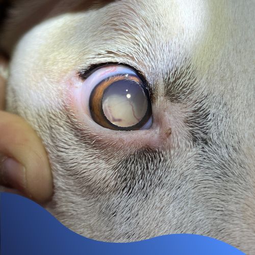
Corneal sequestra are areas of necrotic or dead corneal tissue.
Causes: Often associated with chronic corneal ulceration.
Symptoms:
- Cloudiness
- Opacities
Treatment:
Management of symptoms, severe cases may require surgery such as Diamond Burr.
Corneal Edema
Swelling of the cornea due to fluid accumulation.
Causes: Trauma, inflammation, glaucoma, or endothelial dysfunction.
Symptoms:
- Cloudiness
- Reduced transparency of cornea.
Treatment:
Addressing the underlying cause, including medications and surgery if necessary.
Surgical Intervention Options
Diamond Burr Keratectomy
This surgical technique involves using a specialized tool called a diamond burr to remove or reshape the corneal tissue. It is primarily used in cases where there is corneal pathology, such as superficial corneal lesions, abnormalities, corneal sequestra, and corneal dystrophy.
The Surgery Procedure:
- Anaesthesia – Your dog is placed under general anaesthesia to ensure they are calm and pain-free during the procedure
- Diamond Burr – The vet will use a rotating diamond-tipped burr, which is a small, high-speed instrument, to carefully abrade or remove the affected corneal tissue.
- Wound Management – After the surgery, the cornea is carefully examined and any necessary wound management is performed.
Postoperative Care:
- Your dog may be prescribed antibiotics or anti-inflammatory medication to help with the healing process and prevent infection.
- You will need to closely monitor signs of complications, such as infection or corneal ulceration.
Advantages of Diamond Burr Surgery:
The diamond burr allows for the precise removal of affected tissue, it is also considered as minimally invasive compared to other surgical techniques.
Corneal Grafts (Conjunctival pedicle graft)
This surgical technique involves using a flap of conjunctival tissue to cover and protect the affected area of the cornea.
The Surgery Procedure:
Conjunctival pedicle grafts are often used when other treatment options, such as diamond burr surgery. A conjunctival pedicle graft is a technique that involves using a flap of conjunctival tissue to cover and protect the affected area of the cornea and sutured into place.
Postoperative Care:
- Topical Medications – Your dog is usually prescribed topical antibiotics and anti-inflammatory medications to prevent infection and manage inflammation.
- Monitoring – You will need to closely monitor the surgical site for any complications.
Advantages of Conjunctival Pedicle Grafts:
The procedure promotes healing and the graft maintains its blood supply aiding in the delivery of essential nutrients to the cornea.
If you are concerned your dog is displaying signs of any of the conditions mentioned in this blog, please seek veterinary help by booking a consult.
We hope you have found this information helpful, until next time…

