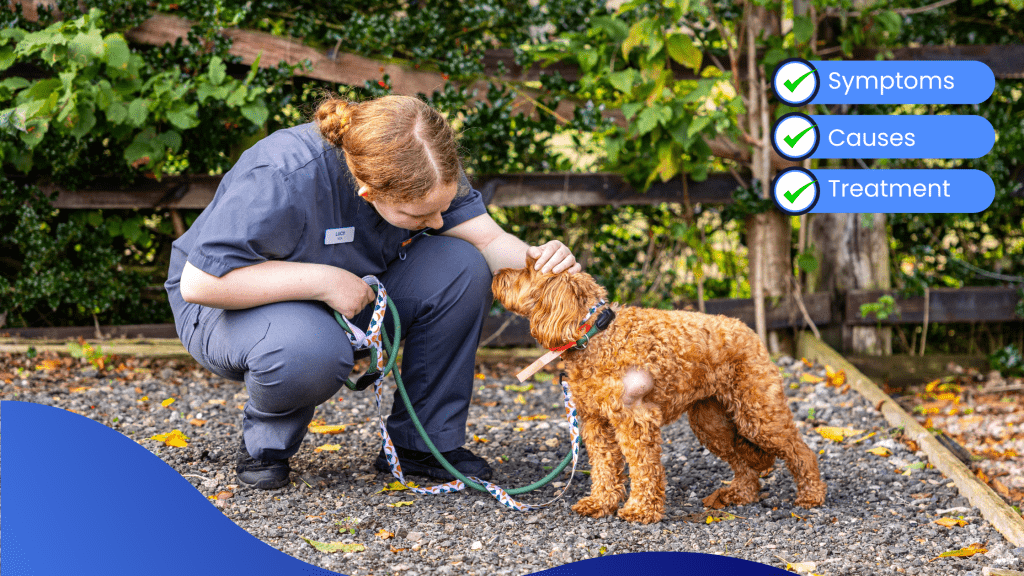
Does your dog have a lump or bump on or under the skin? We know how worrying it is when you find a lump or bump on your dog, but not all lumps are cancerous. There are many types of lumps, early detection is the key to a better chance of recovery, let’s explore the possibilities…
Although bumps and bumps are more common in older dogs, younger dogs can get them too.
Most tumors and bumps are benign (not cancerous), but some can be malignant (cancerous).
The older your dog is, the more likely it is to have melanoma. The good news is that early detection and treatment of cancerous masses can increase the chances of recovery.
How do we diagnose lumps and bumps
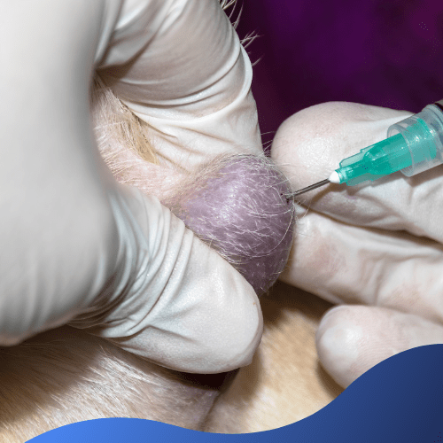
Fine needle aspiration (FNA)
Our veterinarian will first determine if the procedure can be performed during consultations without the use of sedation.
If we determine that we can use this technique for diagnosis, a fine needle will be inserted into the mass to aspirate cells, then placed on a slide.
The slide is then stained and the slide is viewed under a microscope to examine the cells.
We can send the slide to a specialist (pathologist) in the lab for examination.
It is estimated about 95% of tumors and bumps can be diagnosed through FNA.
Impression Smear
If the lump discharges fluid, we may rub a slide into the lump, then stain it and examine the fluid as with an FNA.
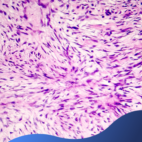

Biopsy
If the FNA isn’t successful in diagnosing the type of lump or bump, or it only determines blood or fluid, taking a biopsy would be the next course of action.
Your dog will receive a sedative or anaesthetic and a small part of the lump or the entire lump will be removed.
The lump is then placed in formalin and sent to the lab, where thin sections of the lump are examined under a microscope.
Lab Test
If the lump contains fluid, the fluid could be sent to the lab to culture and check for infectious agents like fungi or bacteria.

Types Of Lumps and Bumps
Benign (non-cancerous) lumps and bumps
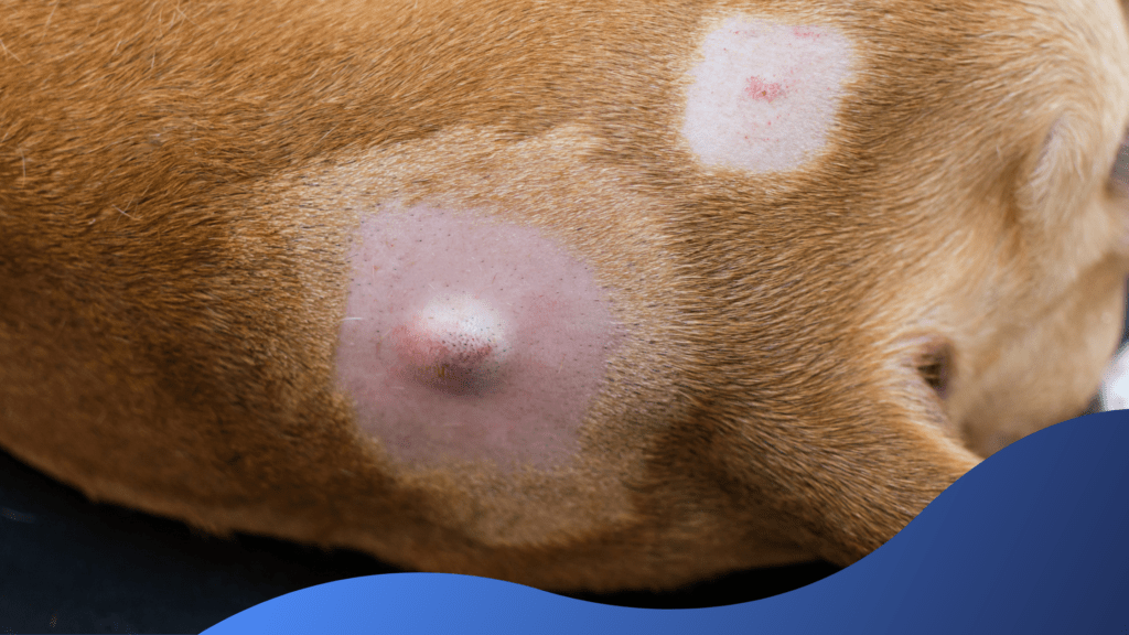
Benign lumps and bumps do not have the ability to invade other tissues and spread to sites outside of their presence. Most are of little concern, but those that continue to grow can cause problems, such as limited movement or breathing due to the size of the tumour, or your dog simply cannot tolerate them and start to scratch the area causing sores and irritation. If benign masses are causing problems, their removal should be considered.
Limpomas
Lipomas are the most common benign tumour that dogs can get. They are usually found under the skin of older dogs and are more common in obese dogs. They tend to be round, soft tumours of fat cells that grow very slowly and rarely spread, so it can take up to six months to see any changes. Lipomas can be easily diagnosed by FNA.
If they become too large or interfere with movement (e.g., grow on the back of the legs or in the armpits), we may recommend removing them..
Abscesses
Abscesses are swollen masses of pus that accumulate under the skin caused by an infection.
They will usually need to be drained under sedation and rinsed thoroughly with a clean antibacterial solution. In some cases, we will prescribe antibiotics if necessary.
Hives (urticaria)
Urticaria in dogs is similar to that in humans – a red, round, itchy, and swollen rash on the skin caused by the skin’s reaction to allergens such as bee stings or contact allergies. They usually go away on their own but sometimes steroids or antihistamines are needed to relieve them.
Sebaceous Cysts
Sebaceous cysts are hard cystic materials under the skin that can form due to blocked sebaceous glands. They appear as swellings with a creamy substance inside. The bumps sometimes become red and painful. They are commonly found in middle-back older dogs and can be diagnosed with FNA. Most of them don’t cause problems, so they’re usually left alone unless they’re infected or make your dog uncomfortable.
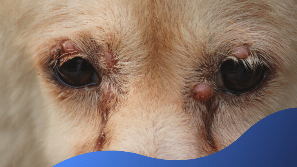
Histiocytomas
Histiocytosis is a sore (or red pimple-like mass) commonly seen in puppies, especially on their limbs.
They usually go away pretty quickly but you should still have them checked by your vet as they can mimic some very nasty cancerous lumps.
Sebaceous Adenomas
Sebaceous adenomas are tumours of the sebaceous glands that appear as multiple wart-like tumors.
They are more common in older dogs with ruffled coats such as Poodles, Maltese, Bichons and their crossbreeds. A biopsy is required for diagnosis, but we can often diagnose these masses just by looking at them due to their classic appearance and slow growth. Most of them don’t cause problems, but sores that irritate the dog or are licked or chewed by the dog should be removed.
Perianal adenomas
Perianal adenomas are tumours that develop around the anus, mainly in older dogs of no gender.
Perianal adenomas appear as slow-growing, non-painful masses around the anus. They usually emerge in the hairless area of the perineum, but technically, they can appear in the prepuce, scrotum, and under the tail. They’re typically superficial and are only rarely adhered to deeper tissues. however, any lump or mass around the anal area should be appropriately evaluated and investigated because malignancies in this area are common.
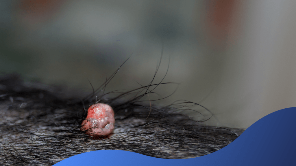
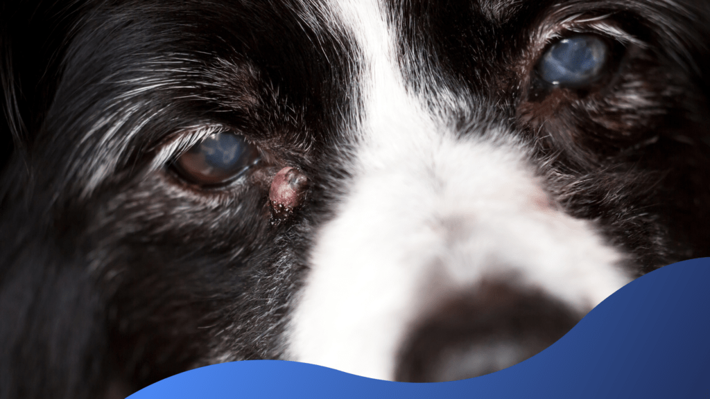
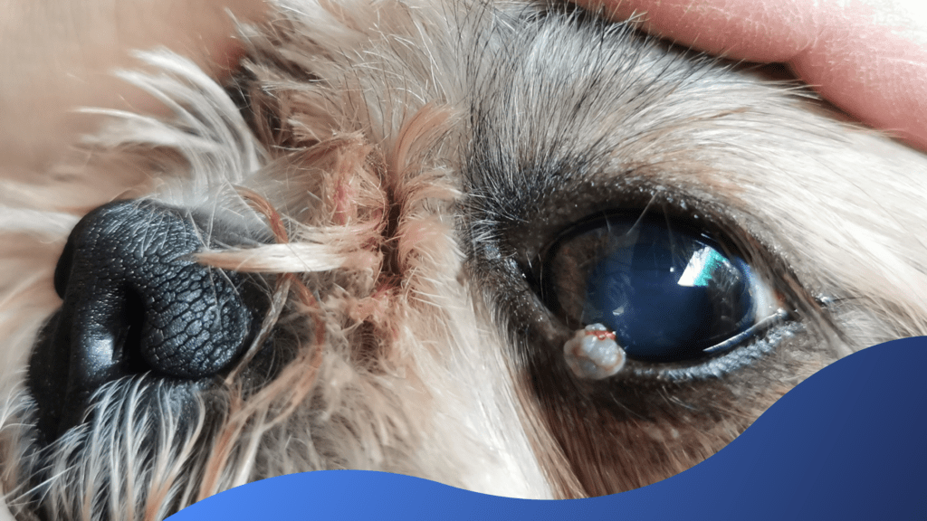
Warts
Warts are more common in puppies, older dogs, and immunocompromised dogs and look like small skin tags or a few small bumps. They are usually found on the head and face and are caused by HPV (papillomavirus). Dogs that go to day-care or dog parks can get warts from close contact with other dogs. A biopsy is necessary for diagnosis, but veterinarians can tell because of their classic fur-like appearance.
No treatment is needed as they usually go away on their own after a few months. However, they can make your dog uncomfortable and if this happens you should consider getting rid of them.
Granulomas
Granulomas can be raised red bumps that may have a crust on the surface or they can be found under the skin and have a firm consistency. They are not usually glued to the muscle. They can look like a very aggressive tumour, so veterinarians often recommend biopsy/resection or FNA. Surgical removal is usually required for treatment.
Haemangiomas
Haemangiomas are tumours of blood vessels or tissues beneath the skin. Sun exposure can lead to their development, but this is not always the case. Diagnosis is made by biopsy or surgical excision with the specimen examined by the pathologist. This is always recommended as these tumours can change over time to become malignant. Surgical excision can be curative if the tumour is benign.
Malignant (cancerous) lumps and bumps
Malignant bumps and tumours develop and can spread throughout the body and affect organs such as the liver and lungs, as well as the brain and bones. They can spread by growing locally (destroying nearby tissue) or by metastasis (tumour cells enter the bloodstream or lymphatic system to spread to other parts of the body). It is important that malignant bumps on your dog are surgically removed as soon as they are diagnosed to prevent them from spreading and causing serious consequences. Chemotherapy and radiation are also often used to stop the spread.
Mast cell tumours
Mast cell tumours are a tumour of the blood cells and of the immune system and account for up to 25% of all tumours. They are more common in dogs over 8 years of age. Mast cell tumors can look like many other tumours, so it’s essential that we diagnoses them correctly. Usually we start with FNA. At diagnosis, it is important to check if the tumour has spread to other organ systems.
Fibrosarcoma’s (soft tissue sarcomas)
Fibrosarcoma’s are locally invasive tumours of the connective tissue of the skin that grow rapidly. They are common in large dog breeds. Biopsies are necessary for diagnosis, as they look like lipomas and can be mistaken for them if FNA is not performed. They are often locally invasive and can be difficult to remove because of the need for rapid and careful resection with a wide incision.
Melanomas
Dog melanoma is not caused by sunlight and is much less malignant than human melanoma. Canine melanoma is a tumour involving the cells that give the skin pigment. They can be benign or malignant and appear as dark, slow-growing lumps on the skin. More aggressive tumours develop on the mouth and legs. They must be removed, but they can recur.
Squamous cell carcinomas
Squamous cell carcinomas are skin cell tumours found on non-pigmented or hairless areas such as the eyelids, vulva, lips, and nose, and appear as raised, raised sores. scales. This is another tumour caused by too much sun exposure.
They usually develop by local invasion and must be removed. If left for too long, they can cause disfigurement and pain, as well as spread to the lymph nodes and other organs, eventually leading to death.
Mammary carcinomas (breast cancer)
Breast carcinoma is a cancerous growth of the mammary glands. They occur more often in female dogs that have not been sexed. Mammary gland tumors can be benign, but it should be noted that mammary tumors in male dogs are usually malignant. They have spread by metastasized to the lymph nodes, mammary glands, and other organs. In most cases, surgical removal of the breast lump and chemotherapy is an option after removal.
Osteosarcomas
Osteoma is the most common bone tumor, especially in large male dogs. They are caused by abnormal bone cell growth, abnormal hormonal stimulation, previous fractures in the area, or genetic factors. They can cause tumors or bumps to form in the bones, often in the extremities, and often spread to the lungs through metastasis. They are diagnosed by biopsy and laboratory tests on bone and skin tissue. They require surgical amputation, which may include amputation of the affected limb.
Chondrosarcomas
Chondrosarcoma is the second most common bone tumor and usually occurs inside the nose. They are also caused by abnormal bone cell growth, abnormal hormonal stimulation, or genetic factors. Biopsy and examination of bone and skin tissue are also necessary for diagnosis.
Treatment of most malignancies requires surgical resection and chemotherapy and/or radiation therapy. When a tumour or tumour is diagnosed as malignant, your veterinarian will take X-rays of other areas and/or an ultrasound of the abdomen to determine if the tumour has metastasized. In certain cases, your veterinarian will recommend a CT scan to pinpoint the exact location of the malignant cells in the body. This is called staging.
How to prevent lumps and bumps?
Not all lumps and bumps can be prevented, however maintaining the health of your dog’s coat is vital to preventing certain skin conditions which cause lumps and bumps. If your dog has a lump or bump, we advise getting seen by a vet straight away. We can tell you if it is dangerous or not, and what is the best method of treatment.
If your dog doesn’t have any lumps or bumps, you should check them regularly to notice any changes. Thread your fingers through their coat and if you feel a lump or bump, take them to the vet for a checkup right away.
The earlier a tumour or bump is diagnosed, the more effective the treatment.
For the whole of March we are offering a free vet check on any pet that has a lump or bump. Give yourself peace of mind and provide your pet with a healthier future.

The free vet check is only valid for pet’s who have any lumps or bumps, health problems outside of this will be charged as a normal consult with the corresponding consult fee.